If You Viewed One Single Protein Using a Microscope
But these models require computer averaging of data from analysis of thousands or even millions of like molecules because it is so difficult to resolve the features of a single particle. Even though each fluorescing molecule still appears as an approximately 200 nanometer.

An Introduction To The Light Microscope Light Microscopy Techniques And Applications Technology Networks
Danev and Baumeister CC BY 40 Proteins under the microscope A new device called the Volta phase plate could improve the.

. Force Spectroscopy of Single Protein Molecules Using an Atomic Force Microscope Citation Details Title. Scientists routinely create models of proteins using X-ray diffraction nuclear magnetic resonance and conventional cryo-electron microscope cryoEM imaging. To explore the prospects for sequencing protein with it measurements of the force and current were performed as two denatured histones which differed by four amino acid residue substitutions were impelled systematically through the sub-nanopore one at a time using an atomic force microscope.
The force measurements revealed that once the denatured protein. Studying Protein Oligomerization using Fluorescence Microscopy. Force Spectroscopy of Single Protein Molecules Using an Atomic Force Microscope.
Each of these methods has advantages and disadvantages. You could infer things about them using X-ray crystallography or measure their pull on tiny probes using atomic force microscopes but not take a direct image. Using a new method coined COLD scientists at the Max Planck Institute for the Science of Light in Erlangen have now visualized protein structures with a resolution of around 5 Å.
The slide will sit directly under the objective lens of the microscope. If you use a microscope to view an unknown cell what structure should you look for to tell you if it is a eukaryotic cell. IHC and IF can be used to detect a single protein in tissues or antibodies can be multiplexed to detect two or more proteins on one slide.
Be notified when an. Step 1 Place your slide on your microscope. Biologists face a similar problem when studying proteins under the microscope as modern imaging techniques can destroy the molecules.
Solution for If a specimen is viewed using a 53 objective in a microscope with a 153 eyepiece how many times has the image been magnified. In a conventional optical microscope objects less than about 200 nanometers apart cannot be distinguished from one another. The trick of the new technique is to control the violet light to activate only a few molecules at a time so that they are statistically likely to be well separated.
The colours also depend on the thickness of the ice. Electron microscopes can reveal the structures of proteins. This clip will hold the slide in place while you view your sample1 X Research sourceStep 2 Look through the lens at your sample.
Request PDF On Feb 1 2017 Elias Amselem and others published Real-Time Single Protein Tracking with Polarization Readout using a Confocal Microscope. To explore the prospects for sequencing protein with it measurements of the force and current were performed as two denatured histones which differed by four amino acid residue substitutions were impelled systematically through the sub-nanopore one at a time using an atomic force microscope. What kind of structures would you observe if you view a single protein using a microscope.
When you start the. To perform this assay you need to expressed one of your target protein fused to. Under the electron microscope -- A 3-D image of an individual protein by Sabin Russell Lawrence Berkeley National Laboratory 3-D images from a single particle A a series of images of an ApoA-1.
The binary protein-protein interaction can be tested in a yeast-two hybrid assay. Please use one of the following formats to cite this article in your essay paper or report. Now graphene the ultra-thin form of carbon has come to the.
Multiplexing is an excellent way to view how proteins are interacting together within the tissue or to view several different cell populations at once. If you viewed one single protein using a microscope you would observe multiple from SCB 203 at LaGuardia Community College CUNY. Push down on the back of the stage clip to raise the clip and allow you to place the slide under it.
Want this question answered. If you examine frozen water through a microscope using polarizing filters and full wave retardation plate you will see colours that are due to the birefringence of ice - its ability to split polarized light into two components that interfere when the rays pass through the ice and polarizing filters. The force measurements revealed that once the denatured protein.
Ice crystals can be made.

From Confocal Fluorescence Microscopy To Super Resolution And Live 3 D Imaging Microscopes Have Microscopy Fluorescence Microscopy Chinese Academy Of Sciences
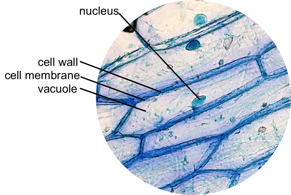
Epidermal Onion Cells Under A Microscope Plant Cells Appear Polygonal From The Cell Diagram Plant Cell Diagram Plant Cell

A Scanning Electron Microscope Image Of A Single Horse Hair Scanning Electron Microscope Images Scanning Electron Microscope Electron Microscope Images
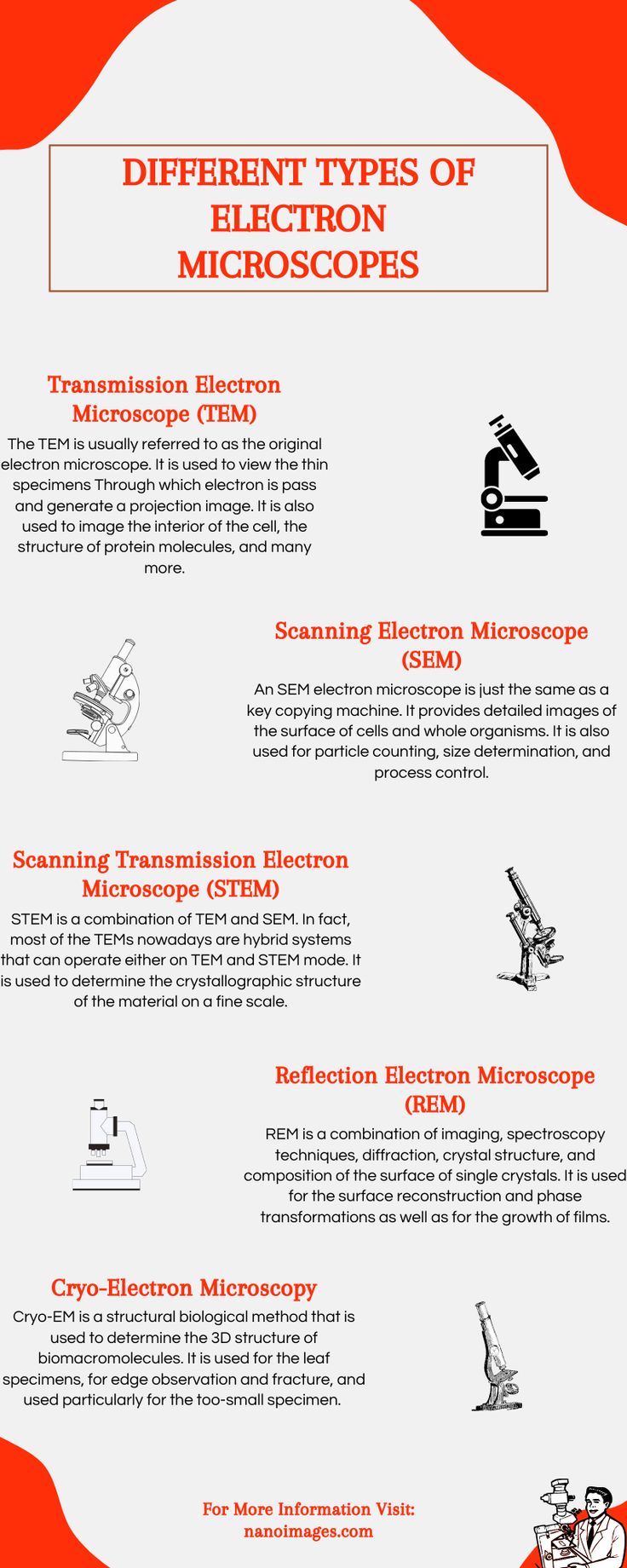
Different Type Of Electron Microscopes Electron Microscope Electrons Scanning Electron Microscope

How To Prepare Microscope Slides Microscope Slides Microscope Slides

30 Piece Microscope Set With 1200x Magnification Magnification Petri Dish Mind Blown
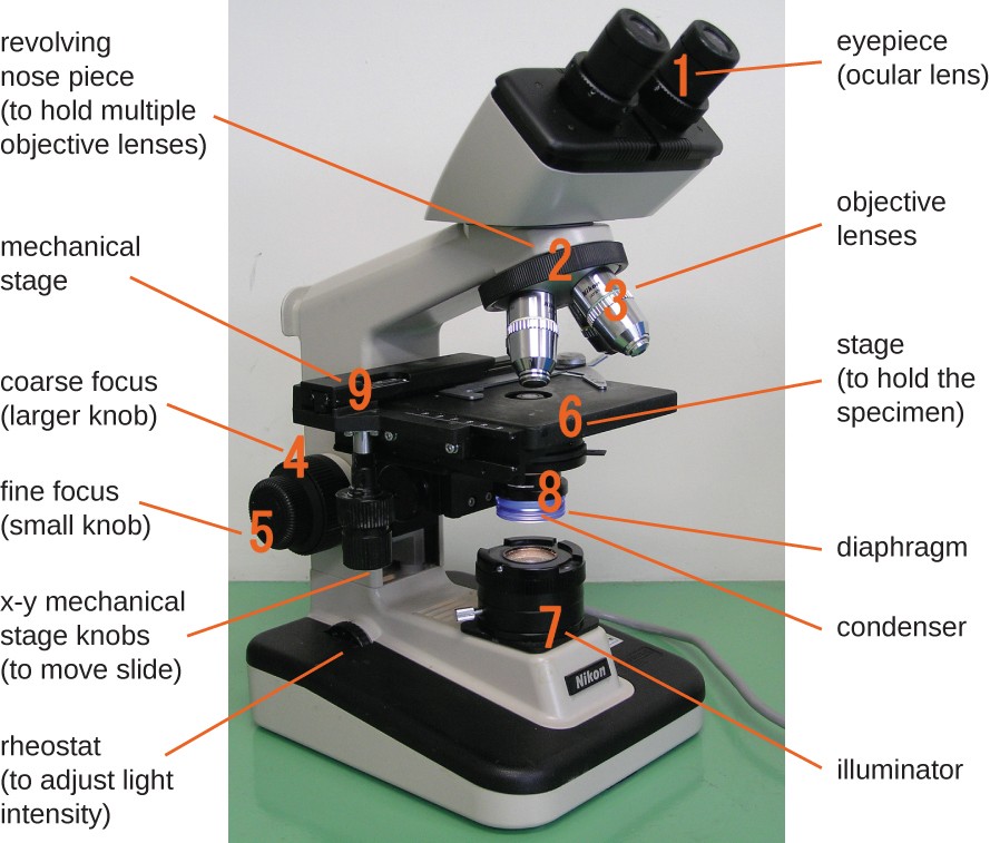
Instruments Of Microscopy Microbiology

Tuscaridium Cygneum Radiolaria Microscopic Photography Science Art Microscopic
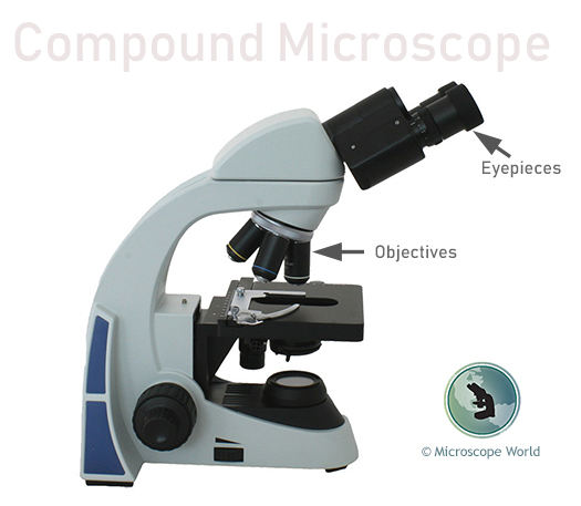
What Is A Compound Microscope Microscope World Blog

Lindsay Langworthy On Twitter Science Biology Solo Taxonomy Membrane Structure

Pin On Teachers Pay Teachers Group Board

Pollen Electron Microscope Microscope Microscopic

Single Yumi 36 Kit Vanilla Overnight Oats Organic Overnight Oats Organic Coconut Sugar

Microscopes Zoom In On Molecules At Last New Scientist

Microscopic View Of Diatoms Microscopic Photography Microscopic Diatom

Pin By Gourav Thakur On My Saves Bullet Journal Journal Save

Cicada Wing Inspired Tech Doesn T Shine Things Under A Microscope Cicada Solar Cell
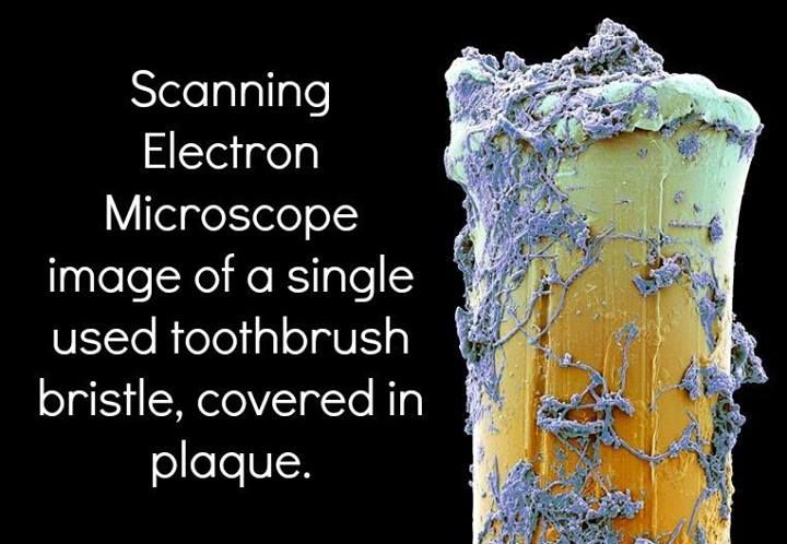
Scanning Electron Microscope Image Of A Single Used Toothbrush Bristle Covered In Plaque Dentist Hygienist Brushing Teeth Toothbrush Bristles Dental Fun
Comments
Post a Comment